
Stem cell technology gives us the possibility to develop mini-organs (termed "organoids") in the lab ("in vitro" as opposed to "in vivo", inside a living organism/animal) [1]. This technology allows us to study how organoids (like mini livers, hearts, eyes and brains) are shaped and develop their functions. They can also be used for drug development, personalized drug testing, and on the longer run for organ transplantations. In collaboration with the largest centre of organoid technology in Norway, Hybrid Technology Hub (HTH)*, we are developing high-content tracking tools and algorithms to guide organoid development. During early embryonic development, retinoic acid generated in a specific region of the embryo serves as an intercellular signalling molecule that guides development of the embryo.
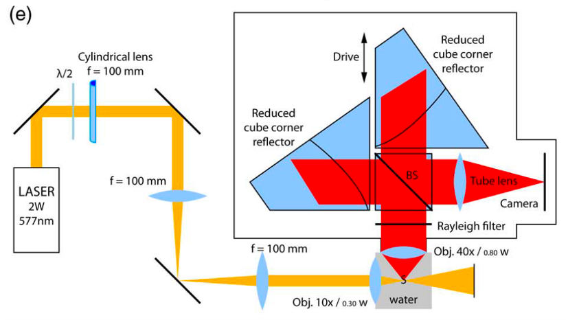
This MSc project is to build a light sheet Raman microscope and use it to image retinoic acid in a developing organoid. The microscope will be built on our existing Open SPIM**. The laser illumination excites molecular vibrations that cause light scattering at various wavelengths that is specific to the vibrational modes of that molecule. In order to identify the molecular fingerprints the detector needs to resolve the spectral intensity in a certain range of wavelengths. This hyperspectral imaging is achieved by imaging repeatedly through an interferometer. The project is to build the interferometer onto the Open SPIM and program the microscope control and image reconstruction and then to use the microscope to image retinoic acid distribution in a developing organoid that our colleagues have prepared. This project will a part of a team with other MSc students, 1 PhD student, 4 postdocs and several faculty from Department of Physics, Chemistry, and HTH. We also have collaboration with the Heintzmann group that have already built a light sheet Raman microscope [2].

[1] M. Hofer og M. P. Lutolf, «Engineering organoids», Nat. Rev. Mater., bd. 0123456789, 2021.
[2] W. Müller, M. Kielhorn, M. Schmitt, J. Popp, og R. Heintzmann, «Light sheet Raman micro-spectroscopy», Optica, bd. 3, nr. 4, s. 452, 2016.
* Hybrid Technology Hub. https://www.med.uio.no/hth/english/
** Open SPIM. https://openspim.org/