
Contact information:
Facility Manager: Kristina Dunkel
Location: Sem Sælands vei 1
0371 OSLO
Norway
Instruments:
- ZEISS Axioscan 7 Microscope Slide Scanner
- three objectives (5x, 10x, 20x)
- cross-polarized light with up to six polarization angles
- Z-stacking
- Two ZEISS Axioscan 5 Petrographic Microscopes
- high-quality camera
- four objectives (5x, 10x, 20x, 50x/100x)
- capturing, processing, and analysis of images in the Zeiss Zen software
Description of services:
- Digitization of thin sections and other microscopic samples
- Acquisition of high-quality, high-resolution scans (pixel size: 0.690-0.173 µm) in brightfield and cross-polarized light with up to six different polarization angles
- Imaging of 3D-objects (e.g., fluid inclusions, palynological samples) by acquiring and combining scans at varying focus heights
- usage of the petrographic microscope
About the Microscope Slide Scanner
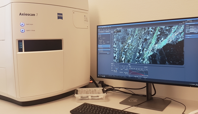
The slide scanner contains an optical microscope with a powerful light source and camera. Slides are automatically loaded onto the microscope stage and moved to different positions, so that many separate micrographs can be taken, which are simultaneously stitched together to produce scans of the whole slide within minutes.
Scans can be acquired in cross-polarised light with different angles of polarization to get a full overview of interference colour, undulose extinction, and other microstructural features.
Three-dimensional objects can be imaged sharply by combining pictures taken at different focal depths (Z-stacking).
Users can view, process, and analyse the scans on their own time using the free version of the Zen software available on the Zeiss website.
Both the Microscope Slide Scanner and the petrographic microscopes are in BookitLab!
The laboratory can by appointment be used for research activities and/or to assist students at the Department of Geosciences.