Contact information:
Facility Manager: Siri Lene Simonsen
Location: Sem Sælands vei 1
0371 OSLO
Norway
Instruments:
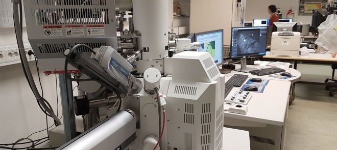
- Hitachi SU5000 FE-SEM (Schottky FEG) including low-vacuum mode and inlens SE-detector (2015). The microscope is equipped;
- Dual Bruker Quantax XFlash 30 EDS system
- Bruker e-Flash HR EBSD system with Argus
- Delmic Sparc Advanced CL system
- Hitachi TM4000Plus equipped with Bruker EDS system (2021).
The laboratory has the following sample preparation equipment:
- Carbon Coater Cressington 208C (shared with the Electron Microprobe Laboratory)
- Qpol Vibro vibratory polisher
Description of services:
- High resolution SE and BSE imaging of rock thin sections and 3D samples
- Low vacuum capabilities for non-coated samples
- Semi quantitative chemical analysis and mapping with EDS
- EBSD and HR EBSD crystal orientation measurements, fully integrated with EDS for simultaneous element analysis
- Cathodoluminescence imaging and spectral analyses
About the Scanning Electron Microscope
The Scanning Electron Microscope (SEM) has a focused electron beam that scans over a surface. The electron beam interacts with the sample creating different signals like secondary electrons (SE), backscattered electrons (BSE), X-rays and light whitch can be recorded with different collectors.
For imaging, the SE, BSE and CL are used. X-rays for EDS (Energy Dispersive Spectroscopy) chemical analyses and element mapping.
The EBSD system reveals the crystal orientation of the minerals providing insight into deformation and microstructure of the sample.
The SEM microscope at the Department of Geosciences is also fitted with a powerful CL system for fast imaging and spectroscopy, giving information of zonation, crystal growth and deformation, which are not seen in SE or BSE.
The laboratory can by appointment be used for research activities and/or to assist students at the Department of Geosciences.
