Principle of TIRF
Total internal reflection fluorescence (TIRF) microscopy (TIRFM) is a powerful microscopy technique that induces excitation of fluorophores within a thin optical section. The method is based on the principle, when excitation light is totally internally reflected due to a refractive index mismatch between the cover glass and cell medium it generates an electromagnetic field, called an evanescent wave. The intensity of the evanescent wave exponentially decays with the distance from the glass surface and only fluorescent molecules within a few hundred nanometers of the glass surface are efficiently excited. Due to the exponential decay of fluorescent signal, images are acquired with a very high signal to noise ratio.
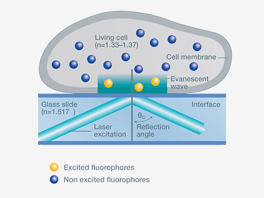
Photo: Olympus-lifescience
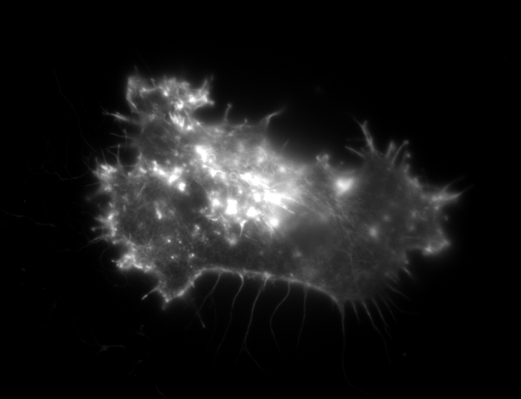
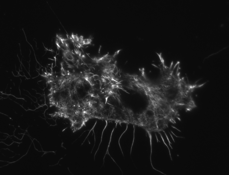
HeLa cell wide field image. HeLa cell TIRF image
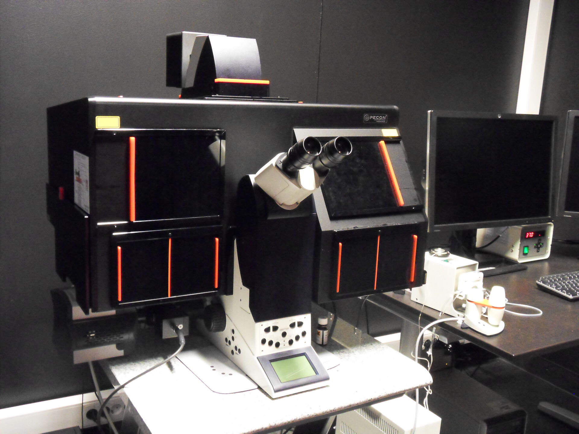
Information about the microscope
Total Internal Reflection Fluorescence (TIRF) microscopes allows efficient detection of fluorescence within a regions close to the sample/glass interface with a high sensitivity, which minimizes the photo damage to living cells. The system offers three wavelengths: 488nm, 561nm, and 635nm; with fast AOTF control and emCCD camera detection. Image acquisition speed can be increased to several images per second using the quad cube filter to avoid repeated mechanical filter changes. Various TIRF slice thicknesses are possible, depending on the excitation wavelength, as well as epifluorescence imaging.
Specifications:
- Heating chamber
- CO2 controller
- Motorized stage
- Autofocus
- Peristaltic perfusion chamber
Laser lines:
- 3 solid state lasers:
- 488 nm
- 561 nm
- 635 nm
Objectives:
| Magnification | Type | Numerical Aperture | Working distance(mm) | Immersion |
|---|---|---|---|---|
| 20x | N Plan | 0.4 | 0.39 | - |
| 40x | HCX PlanApo | 1.25 | 0.1 | Oil |
| 63x | HCX PlanApo | 1.47 | 0.1 | Oil |
| 100x | HCX PlanApo | 1.47 | 0.1 | Oil |
More information from Leica on the TIRF system and objectives.