
Information about the microscope:
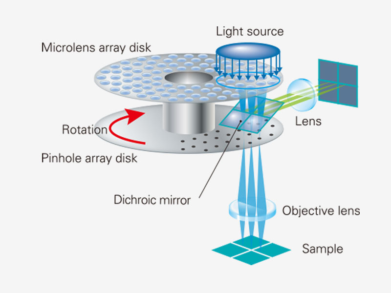
The Olympus SpinSR super resolution imaging system is designed for live cell imaging and the user can switch between super resolution, confocal and widefield imaging. The system is equipped with a Yokogawa CSU-W1 confocal spinning disk unit for fast and sensitive live-cell imaging. A spinning disk confocal microscope simultaneously illuminates many excitation light spots and detects the produced fluorescence from each spot in parallel. A rotating confocal disk scans excitation spots while corresponding pinholes on the intermediate imaging plane reduce the out of focus fluorescence. The spatial distribution of the fluorescence signal is then detected by a camera.
Using the super resolution SoRa disk, detection loss by confocal pinholes is significantly reduced and the SoRa disk provides 3x times brigher super resolution imaging compared to the standard 50um pinhole disk.
Olympus Super Resolution (OSR) technology
The super resolution image is obtained through OSR processing on the acquired image. OSR is a filtering process that amplifies or attenuates a specific spatial frequency component in the image. Reliable imaging with fewer artifacts can be obtained with the optimized amplification factor without spatial frequency shift in the acquired image. The Wiener filter is sometimes used for deconvolution in image processing.
Spesifications:
- Heating chamber
- CO2 controller
- Motorized stage control for multi-location time lapse imaging
- z-drift compensator (IX3-ZDC2), which uses a low phototoxicity infrared laser (wavelenght of 790nm) to identify the sample plane and adjust the focus.
Cameras:
The microscope is equipped with two sCMOS Hamamatsu Orca Fusion cameras with a peak quantum efficiency of 82%.
- Full resolution frame rate 100 f/s
- 2048 x 2048 pixels, 6.5um x 6.5um cell size
Imaging lasers:
- 405nm, 50mW
- 445nm, 75mW
- 488nm, 100mW
- 514nm, 40mW
- 561nm, 100mW
- 640nm, 100mW
Objectives:
| Descrition | Mag | NA | WD | Immersion |
|---|---|---|---|---|
| UPLSAPO | 10x | 0.40 | 3.1mm | Air |
| UPLSAPO | 20x | 0.75 | 0.60mm | Air |
| UPLSAPO | 40x | 1.25 | 0.30 | Silicon oil |
| UPLSAPO | 60x | 1.30 | 0.30mm | Silicon oil |
| PLAPON | 60x | 1.42 | 0.15 | Oil |
| Silicon oil |
FRAP module
The cellFRAP module allowes diffraction-limited laser spot photoactivation and photobleaching and there is only 200us between bleaching an postbleach acqisition. With cellFRAP it is possible to do "click and bleach" to photomanipute a target using direct mouse interaction. Single or multiple points can be bleached or activated with this function. It is also possible to choose a ROI for bleaching before starting the actual experiment.
Bleaching lasers:
- 405nm, 50mW
- 560nm, 100mW